Breast or Mammary gland is a modified sweat gland which is present in both male and female but it remains rudimentary in male whereas well developed in female and thus it is an important accessory organ of the female reproductive system. Breasts symbolizes feminity and beauty and motherhood.
If breast is enlarged in male, this condition is called gynecomastia. This condition may arise due to genetic disorder like Klinefelter’s syndrome and even when excess testosterone is aromatized into estrogen by aromatase enzyme inside the body. Gynecomastia can be treated during initial stage. If its late then, the excess mass removal require surgery. Doctors often prescribe Aromatase inhibitor drugs like anastrozole, letrozole etc which can prevent or reduce gynecomastia in males
Shape
It may be hemispherical, conical, pyriform, pendulum or flat.
Situation
It is located in the superficial fascia of the pectoral region.
It is divided into 4 quadrants i.e upper medial, upper lateral, lower medial and lower lateral.
A small extension of the upper lateral quadrant called the axillary tail of spence (superolateral part) passes through an opening or aperture in the deep fascia and lies in the axilla. This opening is known as foramen of Langer.
Clinical Significance of axillary tail :- It is the site of high occurrence of breast tumor.

Extent
1) Vertically, it extends from 2nd to 6th ribs.
2) Horizontally, it extends from lateral border of sternum to midaxillary line.
Deep Relations
The order of structures from superficial to deep of the breast is as follows :
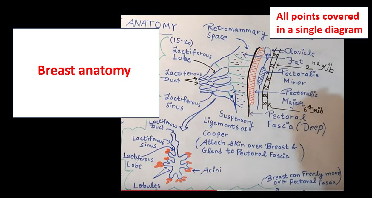
1) The breast lies on the deep fascia (pectoral fascia) which covers the pectoralis major (anterior part).
2) If we go more deeper, there are three muscles associated namely pectoralis major, serratus anterior and external oblique muscle of the abdomen.

Structure
Breast consists of three structures namely skin, parenchyma/mammary gland and stroma.
A) Skin :- Outer covering of the breast and consists of following structures :-
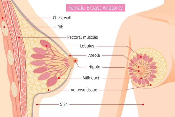
[I] Nipple :-
1) It is conical projection which is present just below the centre of the breast.
2) It is located at 4th intercostal space and 10cm away from the midline.
3) 15-20 lactiferous ducts converge and open onto the nipple.
4) Most sensitive part of the breast as it has many sensory nerve endings.
5) It has circular and longitudinal smooth muscle fibres. Circular smooth muscle fibres make the nipple stiff wheras longituding smooth muscle fibres make the nipple flat.
6) It also has a few modified sweat and sebaceous glands.
[II] Areola :-
1) It surrounds the base of a nipple as a circular pigmented area.
2) It has large number of modified sweat glands (especially at its margin) and few sweat glands and accessory mammary glands.
Modified sebaceous glands produce oily secretion and plays an important role in lubrication of the nipple and areola and prevent them from drying and cracking during lactation.
These glands become enlarged during pregnancy and lactation as small elevations called Montomery’s tubercles.
3) The nipple and areola are devoid of hair and there is no fat surrounding them.
4) Lactiferous sinus lie below the areola where the milk is stored.
B) Parenchyma (Mammary gland) :-
1) It consists of 15-20 lactiferous lobes which converge towards the nipple and open onto it.
2) Each lobe is divided into lobules which consists of a cluster of acini or alveoli and thus mammary gland is also known as compound tubuloalveolar gland.
3) Each lobe is drained by a lactiferous duct. At its termination, each duct has a small expanded or dilation called lactiferous sinus which serves as a reservoir of milk.
The breast is separated from the pectoral fascia by loose areolar tissue called retromammary space and because of this the breast can freely move over the pectoralis major.
C) Stroma :- 1) It forms the supporting framework of the breast.
2) It consists of connective tissue and fat.
3) Connective tissue forms septa called as suspensory ligaments of Cooper. They anchor the skin or dermis to the ducts of the breast and pectoral fascia.
Their atrophy in old age results in the pendulous breast.
4) Fat is distributed all over the breast except the areola and nipple.
Lymphatic Drainage
1) Lymph nodes :-
A) Axillary lymph nodes :-
They lie in the axilla and divided into four groups namely anterior/pectoral,posterior,central and lateral.
B) Internal mammary nodes which lie along the internal thoracic vessels.
C) Supraclavicular nodes which lie above the clavicle.
D) Posterior intercostal nodes
E) Cephalic (deltopectoral) nodes
F) Subdiaphragmatic and Subperitoneal nodes
2) Lymphatic Vessels :-
The drainage of lymphatics is divided into two categories :
1) Superficial lymphatics :-
It drain the skin of the breast except nipple and areola.
2) Deep lymphatics :-
It drain the parenchyma of the breast and also drain nipple and areola.
 Attribution and Credits via wikimedia commons for above image is given here
Lymphatic drainage occurs as follows :-
1) Lymph from lateral quadrants (both upper and lower) of the breast is drained into anterior axillary lymph nodes.
2) Few Lymph vessels from the lower lateral quadrant drain the lymph into posterior intercostal nodes.
3) Lymph from medial quadrants (both upper and lower) of the breast is drained into internal mammary lymph nodes.
4) Few lymph vessels from the lower medial quadrant drain the lymph into subperitoneal lymph plexus.
5) Lymph vessels present deeply in the breast pierce pectoralis major and clavipectoral fascia and drain the lymph into apical group of axillary lymph nodes.
[Important points to Note] :-
1) About 75% of the lymph from the breast drains into the axillary nodes, 20% into the anterior thoracic nodes and 5% into the posterior intercostal nodes.
2) Anterior lymph nodes drain lymph from both the inner half as well as outer half of the breast.
3) A plexus of lymph vessels deep to the areola is known as subareolar plexus of Sappey. Most of the Lymph from the gland and subareolar plexus drain into the anterior lymph nodes.
4) The superficial lymphatics of the breast of one side communicate with those of the other breast and this may result in bilateral malignancy].
Lymph drainage route :-
The lymph from anterior and posterior groups first goes to the central and lateral groups –> Deltopectoral lymph node –> apical group of axillary nodes –> Supraclavicular lymph nodes.